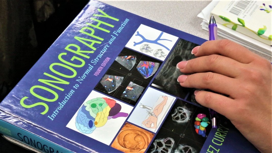


Ultrasound sonography is a non-invasive medical procedure that uses the echoes of high-frequency sound waves (ultrasound) to construct an image (sonogram) of internal organs or body structures. In sonography, a transmitting device (the transducer) sends out high-frequency ultrasound waves. Harmless sound waves, which contain no radiation, bounce off the surfaces of the object they hit. The reflected sound forms an echo which is visualized on the screen.
Education is the passport to the future, for tomorrow belongs to those who prepare for it today.
• Abdomen (AB) – evaluation of all the soft tissues, blood vessels and organs of the abdominal
cavities (for example, liver, spleen, urinary tract and pancreas)
• Breast (BR) – evaluation of breast abnormalities that are found with screening or
diagnostic mammography
• Cardiac (Adult Echocardiography (AE), Pediatric Echocardiography (PE) and Fetal
Echocardiography (FE) – evaluation of the anatomy and hemodynamics (blood flow) of the heart,
its valves and related blood vessels, including Adult Echocardiography (AE), Pediatric
Echocardiography (PE) and Fetal Echocardiography (FE)
• Musculoskeletal (MSK) – evaluation of joints and soft tissue
• Pediatric Sonography (PS) – evaluation of the head, spine, chest, hips/ joints and the male and
female genitourinary system of the pediatric patient
• Obstetrics (OB)/Gynecology – evaluation of the female reproductive system
• Vascular Technology (VT) – evaluation and analysis of the hemodynamics (blood flow) of
peripheral and abdominal blood vessels